A Better Way to Detect and Diagnose Prostate Cancer
 Licking Memorial Hospital (LMH) is using a new procedure to detect and diagnose prostate cancer. The MRI Fusion Prostate Biopsy is an improved method for prostate cancer detection, diagnosis and monitoring. The semi-robotic 3D magnetic resonance imaging (MRI) and ultrasound fusion targeted biopsy combines advances in prostate imaging with traditional, Transrectal Ultrasound (TRUS) Guided Prostate Biopsy for a targeted, more accurate biopsy of the prostate.
Prostate cancer is one of the most common cancers among men. The standard methods for detecting prostate cancer have not changed much in the last 30 years. A digital rectal exam is not very effective because the finger cannot reach the entire prostate, so some cancer could be missed. The prostate specific antigen (PSA) blood test, which detects elevated levels of proteins in the blood, has been a companion to the rectal exam. An elevated PSA level could indicate the presence of prostate cancer.
During a TRUS prostate biopsy, between 12 and 24 needles are inserted into the prostate, guided visually by ultrasound. The ultrasound helps to properly place the needles, but it cannot distinguish between healthy tissue and cancerous tissue, which makes it difficult to get an accurate sampling. Dangerous tumors may be missed while less harmful ones are detected. This can lead to misdiagnosis and unnecessary treatment.
An MRI can reveal details in soft tissue, such as the prostate, more efficiently than an ultrasound. Therefore, it provides a more accurate finding of suspicious areas on the prostate to determine if a biopsy is needed. Because the confines of an MRI machine are not ideal for performing a needle biopsy, a fusion of MRI and ultrasound is beneficial.
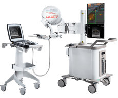
A patient who already has undergone a TRUS Guided Prostate Biopsy with a negative result and continues to have a high PSA level is eligible for the MRI Fusion Prostate Biopsy. The patient will have an MRI of the prostate to detect suspicious areas. A radiologist then will mark those areas on the MRI using specialized software in a process known as “mapping.” A 3D model of the mapping is then exported to be used on LMH’s ARTEMIS Fusion Biopsy System during the day of the surgery.
The MRI Fusion Prostate Biopsy is performed at LMH. Monitored “twilight” anesthesia care, similar to what is used during a colonoscopy, is required for a quality biopsy. The urologist uses ultrasound imaging to acquire more images of the prostate. Those images then are fused with the MRI mapping to create a model of the prostate. The urologist uses a semi-robotic arm to navigate the ultrasound probe and biopsy instrument to target the suspicious locations on the map created by the radiologist. Multiple samples are taken for further examination.
Patients should expect to spend three to four hours at LMH on the day of their appointment. Arriving one hour prior to the procedure is required. The biopsy itself takes approximately 30 minutes to complete. The patient will be monitored in recovery by the nursing staff for about an hour before being discharged. Typically, patients are able to return to work the next day. Most patients will experience blood in their urine for a few days, but it is not unusual for it to last several weeks following the biopsy. Blood also may be present in a bowel movement, which can be resolved with water consumption and rest. Patients also can expect to see blood in their semen for four to six weeks following the biopsy. Patients who become unable to urinate or develop a fever of 100 degrees after the biopsy should call their physician immediately.
| Posted On : 9/17/2020 10:54:37 AM